Tendon Diagram / Hand Tendon And Muscle Anatomy. Learn vocabulary, terms, and more with flashcards, games, and other study tools. Tendons are similar to ligaments; Allows the foot to be turned inward and also supports the arch of the foot. Human hand tendon diagram (page 1) hand tendons diagram muscle blank drawing these pictures of this page are about:human hand tendon diagram this small muscle is located at the top of the shoulder and helps raise the arm away from the body. Tendon, tissue that attaches a muscle to other body parts, usually bones.
You may be able to treat forearm tendonitis with rest and. Pain in tendons between thumb and index finger. The muscle belly then crosses the entire upper arm and separates into two tendons. Related posts of shoulder muscles and tendons diagram anatomy muscles human body video. On the other hand, the insertion is where a tendon attaches that muscle to the *more* movable bone.
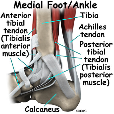
Foot And Ankle Huntsville Al Madison Al Sportsmed Orthopedic Surgery Spine Center from sa1s3optim.patientpop.com Tendons, located at each end of a muscle, attach muscle to bone. Again, our knowledge of how mechanical stimulus mediates ligament and tendon structure is more empirical and less. They propose there are 3 stages to this continuum. To bend the elbow and to turn the palm of the hand towards the sky. The extensor tendon compartments of the wrist are six tunnels which transmit the long extensor tendons from the forearm into the hand. Proper care to be taken to prevent them from injuries. Ligaments join the knee bones and provide stability to the knee: Here you can see the tendons that extend down the top of your foot toward your toes, allowing you to curl your toes upward if need be.
Ligaments and tendons serve similar purposes, but in different ways.
Tendon hand tendons hands feet pinterest and muscles human muscle system human muscle system human muscle. It can be used by a teacher or student for academic purposes. The muscle belly then crosses the entire upper arm and separates into two tendons. Forearm tendonitis is inflammation of the tendons of the forearm. One tendons inserts onto the forearm bone, the radius, and the second spreads out to join the fascia along the upper part of the forearm. Tendons are found throughout the body, from the head and neck all the way down to the feet. The achilles tendon is also called the calcaneal tendon. Brings trunk forward, and aids expiration. Attaches the calf muscles to the calcaneus, most important muscles for running, jumping, walking etc. The changes in ligaments and tendons generally occur more slowly than adaptation in bone, because ligaments and tendons have less vascular supply. They propose there are 3 stages to this continuum. Ligaments and tendons of … category: Proper care to be taken to prevent them from injuries.
The anterior cruciate ligament prevents the femur from sliding backward on the tibia (or the tibia sliding forward on the femur). The achilles tendon is also called the calcaneal tendon. Diagram of knee tendons and ligaments. Tendonitis is the swelling of a tendon, which is a thick cord attaching a muscle to a bone. Tendons that make this possible include:
3 Schematic Representation Of The Hierarchical Organisation Of Collagen Download Scientific Diagram from www.researchgate.net The foot diagram has a complex structure made up of bones, ligaments, muscles, and tendons.understanding the structure of the foot is best done by looking at a foot diagram where the anatomy has been labeled. Connect by text or video with a u.s. A muscle's origin is where a tendon attaches it to the *less* movable bone. 2 ligaments (trapezoid& conoid ligaments) attach the clavicle coracoid process of scapula these tiny ligaments (w/ acominoclavicular joint) keep scapula attached to clavicle. This diagram depicts anatomy of the lower leg achilles tendon. Start studying muscles and tendons. Proper care to be taken to prevent them from injuries. Human hand tendon diagram (page 1) hand tendons diagram muscle blank drawing these pictures of this page are about:human hand tendon diagram this small muscle is located at the top of the shoulder and helps raise the arm away from the body.
Tendons are found throughout the body, from the head and neck all the way down to the feet.
Tendons generally have a very complex structure; Pain in tendons between thumb and index finger. Connect by text or video with a u.s. You may be able to treat forearm tendonitis with rest and. Tendon hand tendons hands feet pinterest and muscles human muscle system human muscle system human muscle. The achilles tendon is the largest. A muscle's origin is where a tendon attaches it to the *less* movable bone. The achilles tendon is a tough band of fibrous tissue that connects the calf muscles to the heel bone (calcaneus). One of the most important tendons in terms of mobility of the leg is the achilles tendon. Diagram of knee tendons and ligaments. It can be used by a teacher or student for academic purposes. Tendons are sometimes confused with ligaments. Start studying muscles and tendons.
Related posts of shoulder muscles and tendons diagram anatomy muscles human body video. A muscle tendon diagram is usually a symbolic illustration of data using visualization approaches. The tendons have 2 functions: Tendonitis is the swelling of a tendon, which is a thick cord attaching a muscle to a bone. The forearm is the part of your arm between the wrist and the elbow.
Ankle Anatomy Eorthopod Com from eorthopod.com The fleshy, thick part of the muscle is called its belly. The current term that is recommended to describe this cohort of patients is 'tendinopathy'. Related posts of foot tendons and ligaments diagram ankle bones anatomy structure. Forearm tendonitis is inflammation of the tendons of the forearm. Ligaments and tendons serve similar purposes, but in different ways. In the back and elsewhere in the body, tendons attach muscles to bones. One of the most important tendons in terms of mobility of the leg is the achilles tendon. Ligaments join the knee bones and provide stability to the knee:
One tendons inserts onto the forearm bone, the radius, and the second spreads out to join the fascia along the upper part of the forearm.
The achilles tendon is the largest. If you would like to learn all the parts of the foot structure, you have come to the right place. Attaches the calf muscles to the calcaneus, most important muscles for running, jumping, walking etc. The achilles tendon is a tough band of fibrous tissue that connects the calf muscles to the heel bone (calcaneus). Related posts of shoulder muscles and tendons diagram anatomy muscles human body video. In the leg muscles diagram above, there are many muscles that make up your legs and support it to move. A muscle tendon diagram is usually a symbolic illustration of data using visualization approaches. Related posts of foot tendons and ligaments diagram ankle bones anatomy structure. One of the most important tendons in terms of mobility of the leg is the achilles tendon. Check out and click on the image to download it. Proper care to be taken to prevent them from injuries. Allows the action of raising the foot. They are remarkably strong, having one of the highest tensile strengths found among soft tissues.
Share :
Post a Comment
for "Tendon Diagram / Hand Tendon And Muscle Anatomy"


Post a Comment for "Tendon Diagram / Hand Tendon And Muscle Anatomy"